"Humans are born with the ability to collect a lot of visual information." Richard Caprioli, director of Stanford Moore Biochemistry and director of the Mass Spectrometry Research Center at Vanderbilt University School of Medicine, said, "We like patterns, we like photos, we can pass a simple photo Get a lot of information. "
In Caprioli's view, this explains why mass spectrometry imaging (MSI) is becoming more popular. In particular, this technology can help histologists to acquire the expertise that would otherwise take years to master. "It uses not the color dimension, but the molecular dimension. But this fact is not so important, as long as the molecular dimension has enough information." He said.
Mass spectrometry imaging technology is like a high-throughput version of immunohistochemistry, but without antibodies. Mass spectrometry imaging technology does not pre-stain tissue sections with special markers. It uses a mass spectrometer to select and draw the spatial arrangement of hundreds of molecules at once. Researchers do not need to know in advance which molecule is more important, they can use this technique to draw and select, and the speed is very fast. "Our instrument has a laser and can do 5000 mass spectrometers per second." Caprioli said that this speed is enough to scan the entire tissue microarray including hundreds of patients' living tissues within an hour.
However, the application of mass spectrometry imaging technology also has obvious obstacles. For example, the image resolution increases as the spot size decreases, but it reduces the yield of ionic materials. This technique does not have a preliminary separation step, so only the most abundant molecules may be extracted. In computing, the challenge for researchers is how to analyze the data, especially how to truly understand the data. But in any case, the researchers are using mass spectrometry imaging technology for research, whether it is to determine the drug metabolites in tissue sections at subcellular resolution, to confirm the biomarkers of disease, or to identify the boundaries of tumors, etc. They are even introducing the technology into the clinic, at least close to clinical research.
Strategies for mass spectrometry
So, what is mass spectrometry imaging technology? Just like a standard digital photo, the color of digital imaging is formed by the superposition of three color channels of red, green and blue, and the color of each small pixel on the screen is composed of these three Consists of the density of colors.
Now, imagine a picture with thousands of color channels. This is the mass spectrometry imaging technique. Caprioli said that each channel is the molecular species or mass spectrum peak you want to display. By covering these different channels with each other, the researchers can produce a color map of the molecular composition and spatial distribution of tissues, whether it is for proteins, neuropeptides, metabolic molecules, or lipids-obviously the demand for lipids is increasing.
Researchers have designed dozens of solutions for mass spectrometry imaging techniques, but as stated in the 2012 review (J. Proteomics, 75: 4883, 2012), only three are the most common. Caprioli's matrix-assisted laser analytical mass spectrometry (MALDI-MSI) scans a matrix-encased tissue slice through an ultraviolet laser raster to create an image. The pixel size of this technology is generally approximately 1 to 10 microns, which means that it can achieve subcellular resolution. However, because it requires the use of MALDI matrix and vacuum environment, MALDI-MSI is not suitable for living samples. At the same time, the matrix is ​​used to absorb the laser energy and transfer it to the sample, but this matrix may be difficult to operate on the sample and generate a large amount of small molecular weight ionization, which will obscure the metabolic region that generates the spectrum.
Nick Winograd, an honorary professor of chemistry at Evan State University of Pennsylvania, adopted the second method—secondary ion mass spectrometry (SIMS). This method ionizes the sample by spraying an ion beam on the surface of the sample (for example, charged C60 molecules or argon cluster beam from Ionoptika, UK) without using a laser. Winograd said that this method has two major advantages. The first is resolution: the pixels obtained by SIMS are about 300 nanometers, while MALDI is only 1 millimeter at best. The other is through deep analysis of molecules. Researchers can use the pits created by collision to “deep dig†this sample and draw its molecular composition in three dimensions.
The third is electrospray desorption ionization technology (DESI). This (non-vacuum) ionization technology uses solvent spray to cover the untreated tissue surface with the solvent and dissolve the molecules on the surface. Then continue to drip the solvent upwards so that the solutes splash onto the mass spectrometer for ionization and analysis. (Prosolia company commercialized the DESI technology, which was co-founded by R. Graham Cooks, the inventor of the technology and Purdue University chemist.)
The three technologies DESI, MALDI, SIMS and their variants all use the so-called "microprobe" mode of Ron Heeren of the AMOLF Institute of the FOM Institute in Amsterdam, and the resolution increases as the pixel size decreases. The problem here is how to maximize the ionization of the sample from the smallest possible spot. But a smaller photoelectricity means fewer ions are detected, not to mention that the imaging time will be longer (because there will be more pixels).
Heeren prefers the "microscopic" mode. This mode allows faster imaging with defocused pixels. Coupled with a pixel detector such as a CCD, it can effectively capture 262144 (512x512) spectra at once.
"It's like a camera," Heeren explained. "That's just one thing. We made molecular flash photos."
Heeren believes that the key to this "mass spectroscope" is the Timepix detector, which is a combination of CCD and time-of-flight mass spectrometer. (Omics2Image, co-founded by Heeren, has a Timepix detector). He explained that most mass spectrometry devices treat the detector as a large pixel, integrating all ion collisions that reach the surface into a single signal. Timepix divides this signal into 262,000 spatially resolved points, so that when detecting molecules on the imaging surface, they can maintain and record their spatial positioning, and the imaging speed is very fast.
How fast? Heeren said that the MALDI-MSI instrument can produce one pixel per second, reaching a resolution of one micron. So in an area of ​​100x100 mm, it takes 2.7 hours to generate 10,000 pixels. But using a quality microscope and Timepix detector, "we can get this information in a second."
There is also a TRIFT SIMS-TOF system for physical electronics on the microscope, and there is also a MALDI technology on it. The Heeren team is currently using this technology to explore physiological changes under osteoarthritis. "We can even confirm that physiological changes in protein levels and lipid metabolism levels and cartilage mineralization can cause the loss of cartilage mechanical strength," he said.
Normal MSI
Compared with MALDI and SIMS, DESI and laser ablation electrospray technology (LAESI, the laser technology introduced by Protea Biosciences), these normal atmospheric pressure ionization technologies have some special advantages. The most obvious advantage is that they do not require sample processing, they can be operated in normal air and do not require vacuum. Therefore, they can be used on living samples and even on patients.
"My own pursuit in my life is to use unprocessed samples for mass spectrometry." This has been Cooks' goal for decades.
As a researcher, Cooks' job is to extract and determine the structure of plant alkaloids. For a long time, the research was very difficult. He only extracted a little "impure alkaloids, and did not make structural progress." Until he met Carl Djerassi who gave a speech from Stanford University. He said that Djerassi took samples of his materials back to the laboratory, collected their mass spectra, and sent the structure back ten days later. "This makes me believe in the power of mass spectrometry," Cooks said. "At the same time, I also found limitations in the extraction methodology."
Since then, he began to get rid of the technical limitations of biologically unmanageable, mass spectrometry, and developed ionization technology under normal pressure, especially DESI. In 2011, a team jointly led by Cooks and Harvard Medical School Nathalie Agar used electrospray analytical ionization mass spectrometry (DESI-MS) to store brain tumor tissue, and used lipid signature test results to help the computer distinguish different forms and histopathological grades. Glioma (a type of brain tumor).
For this analysis, lipids are a quirky choice. Indeed, lipids are helpless for MSI practitioners, but they must get the most value from them. In standard cell analysis, researchers can isolate cell extracts and remove unwanted parts, which often include lipids. But in MSI and other in-situ applications, researchers must know what is in front of them. In front of them are mainly lipids. But fortunately, lipids are not only highly abundant and easy to ionize, but also have a lot of information.
"If you only look at lipids, its tissue characteristics are much better than proteins." Said Zoltán Takáts, a researcher at the Medical Mass Spectrometry Department at Imperial College London.
Recently, Cooks and Agar applied this method to 32 surgical specimens of 5 brain cancer patients undergoing treatment. The system reports the subtype, grade, and part of cancer cells on a pixel-by-pixel basis. Cooks said the data will allow their team to identify areas of different histopathological grades and supplement MRI data when mapping tumor boundaries. He also emphasized that they used the "cheapest" mass spectrometry instrument, Thermo Fisher's single-stage (as opposed to tandem) low-resolution LTQ ion trap.
But Agar also pointed out that this is still a research project, the team can not pass these results to the surgeons in real time, they collect samples in Boston, but the real imaging is in Indiana. Since then, her team has installed Bruker's AMAZon Speed ​​ion trap using DESI technology in the AMIGO operating room of Brigham and Women's Hospital to test brain tumor cases. The operating room is the national center for image-guided treatment in hospitals. Agar said that they will soon develop a breast cancer test, but the team still can not guide the surgeon to really operate. This method must first be verified, "This will eventually need to be verified through clinical trials."
Simplify data analysis
Ultimately, to push MSI into the clinic, it is necessary to cross the mass spectrometer expert and let those who really need to use it master the technology. However, few clinicians can master the subtleties of MSI technology, data processing and informatics, and no one is willing to spend time learning. In Cooks' view, if this technology is "precious and this mass spectrometry technology requires a PhD to master", it will be difficult to promote. "It needs to be fully automated, and the instrument must not be so delicate. It must be reliable and relatively simple." "
For a typical histopathology application, this is not a problem, because this system can be configured as a turnkey box, which can only be opened by a specific biomarker. Non-imaging mass spectrometers have been routinely used in major clinical laboratories around the world, including Bruker ’s MALDI BioTyper and Sequenom ’s MassARRAY. Caprioli wanted to design an MSI similar to a microscope for histologists and pathologists. The instrument is so small that it can even be tucked under the table. The laboratory technician only needs to learn how to prepare samples and operate the machine, and the software can perform the remaining operations.
But more complex applications like biomarker identification are another matter. "The size of the mass imaging technology data set depends on the number of image pixels and the resolution of the mass spectrometer. In recent years, they have undergone tremendous changes." Lawrence Berkeley National Laboratory scientist Ben Bowen said that he invented mass imaging Technical data analysis software.
As the resolution increases, the pixels shrink. At the same time, researchers conducting "discovery mode" experiments do not know which molecule is more important in advance, so they have to take all molecules into account and make pairwise comparisons on thousands of color channels.
The sum of all these pixels is amazing. Bowen said that his colleague Trent Northen uses mass spectrometry imaging technology in his work and has collected millions of megabytes of data over the years. For beginners, opening data files is a problem, which makes them very dependent on experts who are more proficient in this technology. "You will know why it makes these scientists so unhappy." Bowen said.
To alleviate their burden, Northen and Bowen and Berkeley Lab data visualization expert Oliver Ruebel jointly developed the OpenMSI cloud computing platform. Users can browse and operate the cloud computing data of mass spectrometry imaging technology directly on the browser. According to Bowen, the US Department of Energy ’s National Energy Research and Scientific Computer Center (NERSC) supercomputer is used to support the system, reducing data processing time from days to minutes.
Bowen said that one of his and Northen collaborators can use OpenMSI to study the 50-gigabyte data set in detail. This data set was collected a year and a half ago, but there has been no way to study it. "Now he uses this technology in the (Google) browser." He gave examples, including browsing RGB images, examining the spectrum below, and sharing data with colleagues, "All the functions you can think of in the 21st century Internet , We can all realize this function of mass spectrometry imaging technology on OpenMSI. "
Operating room mass spectrometer
However, to optimize clinical translatability, researchers may have to break away from the imaging part of MSI. This is the research result of Takáts at Imperial College London.
Takáts was a postdoctoral fellow before Cooks and described DESI for the first time as the first author of the paper. He has developed and is testing a new non-vacuum ionization technology-Rapid Vapor Ionization Mass Spectrometer (REIMS), and designed the iKnife smart scalpel. Surgeons can handle tissue histology and histopathology in the operating room.
"The final device is very simple," Takáts explained, and it also relies on electrosurgical techniques, which use current to vaporize tissue. The smoke released in this process is a combination of tar, particulate matter, and ionized lipids. IKnife continuously extracts the sample into a Teflon tube attached to it, and then puts it into the mass spectrometer.
Over the past few years, the database established by Takáts includes nearly 200,000 lipid samples from human cancer and healthy tissues. Through these data, he confirmed that different samples can be distinguished by lipid biomarkers. Therefore, using the ionized lipid profile generated during electrosurgery, his system can determine in real time whether the tissue under iKnife is healthy or cancerous, and its histological status.
But it should be noted that there is no imaging. "The diagnosis is a histological identification." Takáts said, "This system will tell you that it is non-small cell lung cancer, stage 2 or something."
In Hungary, Germany and the United Kingdom, iKnife (the research and development results of MediMass and Imperial College) has been tested in more than 500 operations. "In most cases, 100% classification accuracy can be achieved." Gastrointestinal cancer, liver cancer, lung cancer, breast cancer and brain cancer. In some cases, doctors thought it was a tumor, but the technology proved to be only benign tissue or inflammatory disease. Now, Takáts is developing a new system that can perform similar evaluations for endoscopy.
Takáts said that in the end, this application may make MSI "significant", not only as a research tool, but also as a routine clinical technology. He pointed out that histopathology researchers may be reluctant to accept this relatively slow and expensive technology. But he also said that the speed and price of this instrument will be improved. In the past, the best time to perform this test was to wait for half an hour after the dissection before performing the test. If this technology can provide a diagnosis in a few seconds by the doctor and is in vivo, then people will be more inclined to this technology.
"The advantages of this system are beyond histopathology," he said.
Gear Clock On Wall is a fashion item for both time and decorative function. You can see the gear moving and every gear has its own meaning, whatever hour, minute or seconds, as you can see the time going. Special Gear Clock not only including the table Gear Clock, but also the Wall Gear Clock. But hanging a gear clock on wall is like hanging a art work to increase the sense of art of home.
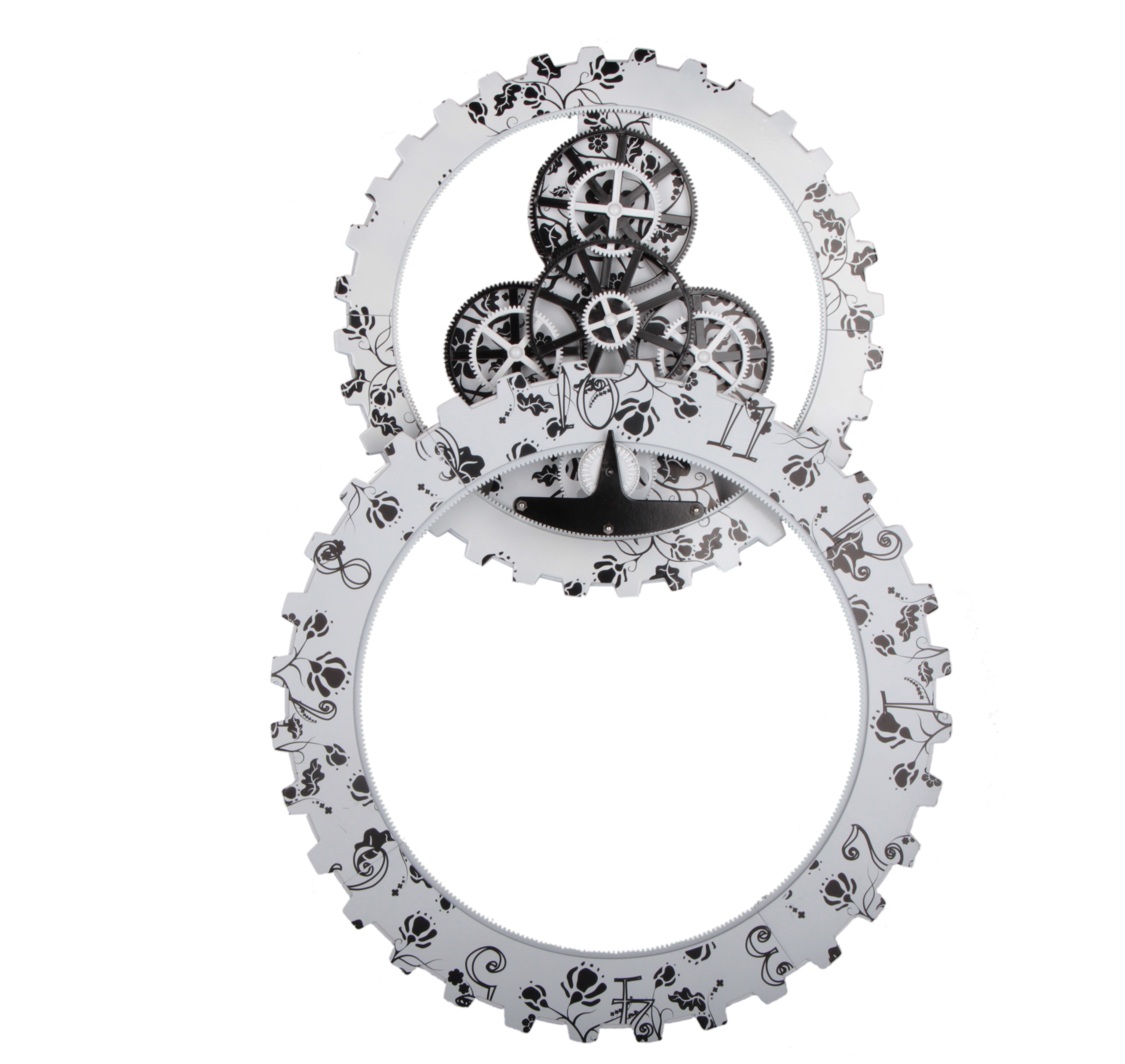
Gear Clock On Wall
Wall Clocks At Target, Modern Wall Clocks, Big Gear Clock,Gear Clock Design,Gear Clocks For Sale
Guangzhou Huan Yu Clocking Technologies Co., Ltd. , https://www.mk-times.com